home
| people
| publications
| software
| SVD-PCA
| q-bio
| book
| cv
Structural Biology
We are interested in using protein structure to infer protein function
(Protein Function
Inference) and in small-angle scattering methods for determining
large-scale shape changes in proteins (Small-Angle
Scattering).
Protein Function Inference
The ongoing determination of genomic DNA sequences raises questions concerning the biological function of each gene. To address these questions, we are developing methods for inferring protein function based on analysis of protein sequence and structure. We have developed a method called Dynamics Perturbation Analysis (DPA) that uses analysis of protein dynamics to predict protein functional sites (Ming and Wall, Proteins 2005, JMB 2006). Similar methods were applied to increase the accuracy of coarse-grained models of protein dynamics, and to study allosteric effects in proteins (Ming and Wall, PRL 2005). Additional projects include estimation of protein configurational entropy, and machine learning prediction of enzyme function. Older related work includes development of
SOESA software (available here) for using a database of protein structures to (read the paper).
Small-Angle Scattering
Proteins and RNA undergo intricate motions as they carry out functions in biological systems. These motions frequently entail large-scale conformational changes that induce changes in the surface structure, or shape, of a molecule. Small-angle scattering is a powerful way to detect such changes, and to study the molecular mechanisms of protein protein function. In collaboration with Jill Trewhella, we have been preforming small-angle scattering studies of second-messenger signaling proteins, most recently cGMP-dependent protein kinase (read the paper). We have also been developing a web portal called SASDAP (see our internal site) for analysis of biomolecules studied using small-angle X-ray and neutron scattering.
Diffuse X-Ray Scattering
This is not an active research area in our lab but please feel free
to contact me for more information about the topic. Diffuse X-ray
scattering studies are
complementary to traditional x-ray crystallography, where an ordered
crystal
of more than a billion molecules is exposed to a beam of x-rays. The
order in the crystal causes the x-rays to be scattered into only
particular directions, called Bragg reflections.
By measuring the number
of x-rays in each Bragg reflection, the molecular structure of
a single molecule can be resolved.
If the molecules in the crystal are moving, the resultant disorder
causes x-rays to be scattered into angles which do not satisfy the
Bragg condition. By measuring this diffuse scattering, the
motions of these molecules can be characterized. Diffuse scattering
is usually thrown away in most crystallographic studies, but advances
in x-ray sources, detectors, and computing have made it easier to use
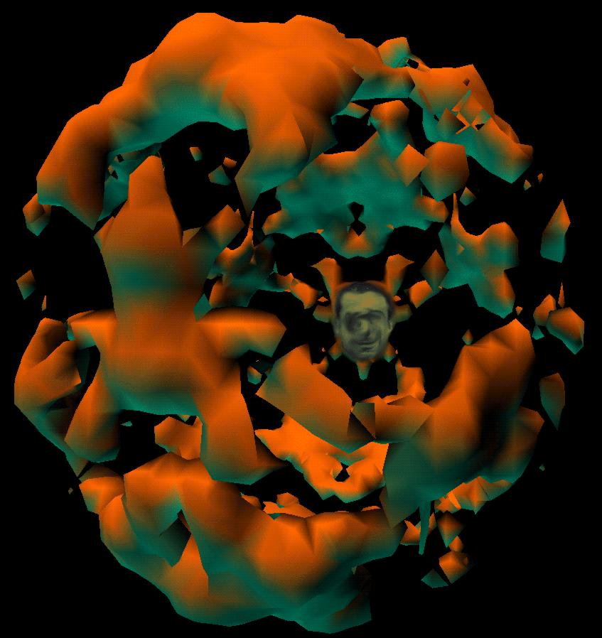
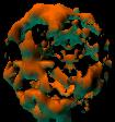
it fruitfully. Wall, Ealick and Gruner
(1997) describes the measurement and characterization of the
diffuse X-ray scattering from crystals of Staphylococcal nuclease,
which produced surprising views of reciprocal space such as that
visible at the right. More details of this study, including detailed calculations of
diffuse scattering based on many kinds of disorder, are described in my thesis,
which can be downloaded here as a PDF file.
The methods were used in a study of dynamics in calmodulin crystals
(see Wall,
Clarage and Phillips 1997).
Michael Wall / mewall@lanl.gov
Los Alamos National Laboratory,
CCS-3 MS B256,
Los Alamos NM 87545
tel: 505.665.4209 / fax: 505.667.1126
Last modified Monday, 29-Jun-2009 16:53:34 MDT